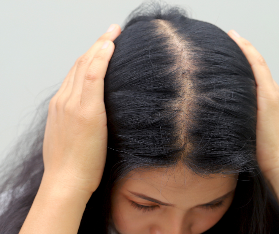
It starts subtly — a wider parting, a slightly thinner ponytail, a moment of shock when the light catches your crown in the mirror. Female crown thinning is one of the most emotionally distressing hormonal symptoms there is, and one of the most inadequately addressed. The answer is not a better shampoo.
Crown thinning in women — clinically known as female pattern hair loss (FPHL) or androgenetic alopecia — is the most common form of hair loss in women, affecting an estimated 40 percent of women by age 50. But it is increasingly showing up in women in their twenties and thirties, and in those cases it is almost always driven by a correctable hormonal or nutritional imbalance rather than by irreversible genetic fate.
Functional medicine approaches female hair loss differently from conventional dermatology. Rather than recommending minoxidil as a first-line intervention and leaving the cause uninvestigated, a functional approach maps the specific hormonal, metabolic, and nutritional drivers behind the hair cycle disruption — and addresses them systematically. This article explains exactly what those drivers are, why the crown is specifically affected, and what a targeted intervention looks like.
Why hair thins at the crown specifically
The distribution of hair loss in women with FPHL — predominantly at the crown and along the central parting, with relative preservation of the frontal hairline — is not random. It reflects the distribution of androgen receptors in the scalp. Hair follicles at the crown have a significantly higher density of androgen receptors, particularly receptors sensitive to DHT (dihydrotestosterone), compared to follicles at the sides and back of the scalp. When DHT levels are elevated, or when crown follicles are unusually sensitive to DHT even at normal levels, the follicles in this region miniaturise progressively — shrinking from thick terminal hairs to fine, short vellus hairs over successive hair cycles.
This miniaturisation is the hallmark of androgenetic alopecia. It is not that the follicle dies — it is that it progressively loses the ability to produce a full-sized hair. The earlier this process is identified and the underlying drivers addressed, the more of this miniaturisation is reversible.
The hair growth cycle and where it goes wrong
In healthy hair cycling, approximately 85 to 90 percent of scalp hairs are in the anagen (growth) phase at any given time, with the remainder in the transitional or resting phases. The most common hormonal drivers of crown thinning work by either shortening the anagen phase, pushing hairs prematurely into the telogen (resting) phase, or causing follicle miniaturisation through DHT-mediated mechanisms. Each of these pathways has a different hormonal root cause.
Crown thinning in women is not inevitable and it is not purely genetic. In most cases it reflects a specific, identifiable hormonal and nutritional imbalance — and the earlier it is investigated, the more reversible it is.
The main hormonal and nutritional causes of female crown thinning
DHT and androgen sensitivity: the primary mechanism
DHT — dihydrotestosterone — is produced from testosterone by the enzyme 5-alpha reductase. In crown scalp follicles with high androgen receptor density, DHT binds to follicle receptors and progressively shortens the anagen growth phase with each successive hair cycle. Over time, the follicle produces thinner, shorter hairs until it is effectively dormant.
In women, DHT-driven crown thinning does not require abnormally high testosterone. It can occur in the context of normal testosterone levels when 5-alpha reductase activity is elevated, when SHBG (sex hormone binding globulin) is low — increasing free testosterone available for conversion — or when the follicle receptors themselves are highly sensitive. This is why a standard testosterone blood test often reads “normal” in women with significant androgenetic crown thinning — the problem is at the follicle level, not simply in the circulation.
Insulin resistance is the single most significant modifiable driver of elevated androgen activity in women — by reducing SHBG and stimulating ovarian testosterone production, it increases the androgenic load reaching the scalp. PCOS is the most common clinical presentation of this pattern, but insulin-driven androgen excess causing crown thinning occurs broadly in women without a formal PCOS diagnosis.
Thyroid dysfunction and hair loss
The thyroid is one of the most powerful regulators of the hair growth cycle. Thyroid hormone receptors are present directly in hair follicle cells, and thyroid hormone is required to maintain follicles in the anagen phase. Both hypothyroidism and hyperthyroidism disrupt the hair cycle, but hypothyroidism is by far the more common driver of crown thinning in women — producing a diffuse, often crown-predominant thinning that is frequently mistaken for genetic hair loss.
Subclinical hypothyroidism and Hashimoto’s thyroiditis are significantly underdiagnosed in South African women. Hair loss is often one of the earliest presenting symptoms — appearing months before TSH rises enough to trigger conventional treatment thresholds. A full functional thyroid panel (TSH, free T3, free T4, reverse T3, TPO and thyroglobulin antibodies) is essential in any woman presenting with new or worsening crown thinning.
Hair loss is one of the first symptoms of thyroid dysfunction — and one of the last to resolve after treatment begins. Investigating and correcting thyroid function early is the single most important intervention for thyroid-related crown thinning.
Iron deficiency and ferritin: the most overlooked correctable cause
Low ferritin — the iron storage protein — is one of the most common and most reversible causes of female hair thinning, yet it is routinely missed because haemoglobin remains normal even when ferritin is critically depleted. The hair follicle is one of the most metabolically active structures in the body, and ferritin is required for the enzymatic reactions that sustain the anagen growth phase. When ferritin falls below approximately 40 to 70 mcg/L, the hair cycle shortens measurably and diffuse thinning — often worst at the crown — begins.
In South African women of reproductive age, low ferritin is extremely common — driven by menstrual blood loss, low dietary iron intake, poor iron absorption from gut dysbiosis, and chronic inflammation that sequesters iron in storage. Many women have been told their iron is “fine” based on haemoglobin alone, while their ferritin has been sitting at levels that are actively driving hair loss for years. Replenishing ferritin to optimal levels — not just the lower limit of the reference range — is frequently one of the most transformative interventions for female crown thinning. Hemagenics by Metagenics provides comprehensive iron and haematinic support formulated for optimal absorption and tolerability.
Cortisol, stress, and telogen effluvium
Chronic cortisol elevation drives hair loss through two distinct mechanisms. First, cortisol directly shortens the anagen phase and accelerates the shift of follicles into the telogen resting phase — a process called telogen effluvium, which produces the sudden, diffuse shedding that many women experience after a period of significant stress, illness, or major life disruption. Because telogen hairs shed three months after the triggering event, many women cannot identify the cause — the shedding seems to appear from nowhere.
Second, chronic cortisol elevation drives adrenal androgen production (DHEA and androstenedione), which feeds into the DHT-mediated follicle miniaturisation pathway at the crown. Women with HPA axis dysregulation therefore experience both telogen effluvium from the cortisol effect and androgenetic miniaturisation from the androgen effect simultaneously — making stress-driven hair loss particularly complex and persistent without proper hormonal investigation.
Oestrogen, progesterone, and the protective hair effect
Oestrogen and progesterone both protect and prolong the anagen phase of the hair cycle. This is why many women experience their best hair growth during pregnancy — when both hormones are at their peak — followed by significant postpartum shedding as they drop. The same mechanism operates in perimenopause, when declining oestrogen and progesterone remove their anagen-sustaining effect and crown thinning often accelerates noticeably.
Progesterone also inhibits 5-alpha reductase — reducing the conversion of testosterone to DHT at the scalp level. When progesterone falls, DHT activity at the crown increases even without any change in testosterone levels. This is why crown thinning frequently worsens perimenstrually and during perimenopause — the same progesterone decline that drives mood and sleep symptoms is simultaneously removing the follicle’s DHT protection.
Targeted functional medicine support for female crown thinning
What a proper investigation looks like
A functional medicine workup for female crown thinning includes a full thyroid panel, sex hormone assessment (testosterone, free testosterone, DHEA-S, SHBG, oestradiol, progesterone), fasting insulin and glucose, a full iron panel including ferritin, vitamin D, zinc, B12, folate, and inflammatory markers. Scalp biopsy may be indicated in cases where the pattern is atypical or where rapid progression warrants ruling out scarring alopecia.
The interventions that follow are highly dependent on which drivers are identified. DHT-driven miniaturisation requires a different approach to iron-deficiency thinning, which in turn requires a different approach to cortisol-driven telogen effluvium. Treating all three as the same condition — as often happens with generic hair loss supplements — produces inconsistent and often disappointing results.
The bottom line
Female crown thinning is almost never purely genetic fate. In the majority of cases it reflects a specific, identifiable, and at least partially reversible hormonal and nutritional imbalance — whether that is DHT sensitivity driven by insulin resistance, thyroid dysfunction slowing the hair cycle, ferritin depletion shortening anagen, cortisol excess driving shedding and androgen production, or progesterone decline removing the follicle’s natural DHT protection.
The first step is understanding your hormonal picture. Take the free hormone assessment quiz at Hormone Reset to identify which imbalance pattern is most likely contributing to your hair loss — and to get clarity on where your investigation and treatment should begin.
Hair is not a vanity concern. It is a visible marker of internal hormonal and nutritional health — and when it starts to go, it is worth taking seriously and investigating properly.
Ready to identify what’s driving your crown thinning and address it from the root?
Take the free hormone assessment quiz